

Observing how new blood vessels are formed in living organisms – and especially in the heart – is challenging in mammals. Therefore, you only ever see endpoints, in other words, that new vessels have formed and which cells they comprise. However, little is known about the actual process of vessel formation, and yet in the future this knowledge could contribute towards repairing tissue damage that occurs as a result of ischaemia, for example, in diabetics or after myocardial infarction.
DZHK scientist Professor Dr. Stefanie Dimmeler and her colleagues at the Institute of Cardiovascular Regeneration of the Goethe-University Frankfurt am Main have thus examined individual cells of the inner layer of blood vessels, the endothelial cells, during development and after tissue damage in a so-called Confetti model. In this model, the scientists can label and distinguish specific cell types using fluorescent proteins. Only endothelial cells fluoresced in three different colours. Since fluorescence is retained when cells divide, individual endothelial cells and their “descendants” can be tracked. With this method, the scientists wanted to determine whether cell division in the formation of new blood vessels is either random, as seen in zebrafish, or whether certain cells repeatedly divide, thus forming the new vessels.
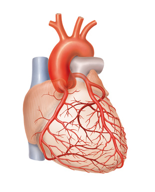
Clonal expansion occurs after myocardial infarction
The researchers could observe that certain cells had divided very frequently in damaged heart tissue after a myocardial infarction. They also determined this cell division known as clonal expansion in skeletal muscle tissue damaged by ischaemia. To this end, the scientists analysed the fluorescence of endothelial cells in tissue sections of the damaged areas. For the researchers, the rate of clonally expanding cells was surprisingly high at 30 to 50 percent. “Yet we might still be underestimating the rate of the observed clonal expansion”, suspects Dimmeler. “For we did not carry out a three-dimensional analysis, but determined the luminous cells in two-dimensional tissue sections.” Further experiments also showed that the vessels formed by clonal expansion are indeed perfused and are thus functional.
In new-born mice, however, the DZHK scientist and her team were unable to determine clonal expansion during retinal angiogenesis. During normal development, the formation of blood vessels thus seems to consist of the random division and integration of cells. This result coincides with the observations in zebrafish, in which angiogenesis during development is also determined by what is known as cell mixing.
Profiling cells
The researchers wanted to characterise the actively dividing cells in more detail and thus analysed which genes are read in individual clonally expanding endothelial cells. “Surprisingly, we found lots of gene products that are typical for the transition of an endothelial cell to a mesenchymal cell”, states Dimmeler. This transition, which is called the EndMT process, is part of many pathological processes, for example, in scar formation or atherosclerosis. Yet in endothelial cells, the gene products typical of EndMT do not reflect a transition, but presumably only an intermediate stage which allows the cells to detach from the cell group to clone.
Clonal expansion as possible myocardial infarction therapy
Dimmeler and her team now want to learn what happens with the clonally expanding cells long-term, because they are currently only able to follow their fate for about two months. “We want to know what will have happened to these cells a year later and whether the new blood vessels are just as good long-term as the old ones”, says Dimmeler.
What clonal expansion is like in elderly patients is also an extremely exciting question to the DZHK scientist. “Perhaps clonal expansion in the elderly is no longer as efficient, which is why after a myocardial infarction, a lot of damaged tissue mortifies and scars, and can no longer be reactivated with the formation of new blood vessels,” says Dimmeler. “If we characterise the clonally expanding cells in more detail, we hope to find ways to re-initiate this process in a targeted manner.”
Original study:
Clonal Expansion of Endothelial Cells Contributes to Ischemia-Induced Neovascularization. Manavski, Y., Lucas, T., Glaser, S. F., Dorsheimer, L., Gunther, S., Braun, T., Rieger, M. A., Zeiher, A. M., Boon, R. A. & Dimmeler, S. Circulation research 122, 670-677, (2018). DOI: 10.1161/CIRCRESAHA.117.312310 .
Contact:
Christine Vollgraf, Public Relations Officer, German Centre for Cardiovascular Research (DZHK), phone: +49 30 3456 529 02, presse(at)dzhk.de
Prof. Dr. Stefanie Dimmeler, Institute of Cardiovascular Regeneration, Institute for Molecular Medicine, Goethe-University Frankfurt am Main, dimmeler(at)em.uni-frankfurt.de