
Diastolic heart failure, also called heart failure with preserved ejection fraction (HFpEF), is a disease that appears contradictory at first glance. Patients exhibit the symptoms of systolic heart failure; for example, they suffer from dyspnoea and water retention. Yet unlike with systolic heart failure, the pumping power of the heart is not impaired. Rather, the problem with HFpEF is that the left ventricle is stiff and does not sufficiently fill with blood. This is also where the terms diastolic and systolic heart failure are derived from. Diastolic denotes the phase when the heart expands and fills with blood, while systolic denotes the phase when the heart contracts and ejects blood.
Whether the heart is insufficiently filled with blood is hard to measure with the non-invasive methods currently used, such as an ultrasound examination, and is often only identified late. Invasive methods are indeed more exact, but expensive, onerous, and they cannot identify the causes of the disease.
New standard for the diagnosis of HFpEF
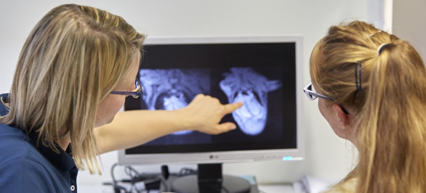
The multicentre study DECIPHER HFpEF-DZHK12 headed by Professor Eike Nagel of the University Hospital Frankfurt aims to establish cardiovascular magnetic resonance imaging (CMR) of the heart as the standard method for diagnosing HFpEF. To this end, the study compares the CMR data with the results of the present gold standard in the diagnosis of HFpEF, invasive haemodynamics, where patients are examined using a cardiac catheter. In order to obtain a comprehensive picture of the validity of CMR, Nagel and his colleagues are also comparing the CMR measurements to the results of ultrasound examinations and analyses of myocardial tissue samples.
A disease with many causes
Using these data, the researchers aim to investigate whether CMR can help clarify why the disease arises, especially since the underlying cause of diastolic heart failure can vary greatly. For example, it can be caused by an inflammation of the heart muscle (myocardium), thickening of the myocardium, or decreased blood flow to the myocardium due to changes to the smallest blood vessels. CMR procedures developed in recent years enable these parameters to be assessed and are for the first time merged in a combined examination in the study. This is because, unlike an ultrasound examination, these new procedures can not only measure the flow and filling of the ventricle, but also see, whether the heart muscle is inflamed, whether there is a pathological proliferation of the connective tissue, or whether there is a change to the small vessels.
“Only once the causes are clear can patients receive targeted medicinal therapy, because the treatment of myocardial inflammation differs entirely to that of a circulatory disorder”, explains Nagel. “Significant therapeutic studies are only possible once we can distinguish between the various patient subgroups.” DECIPHER HFpEF-DZHK12 aims to lay the foundation for this. It is the first multicentre study on the diagnosis of HFpEF worldwide. Beside Frankfurt am Main, the DZHK sites in Berlin, Heidelberg, Göttingen, and Bad Nauheim are also participating. If the results are positive, the study would lead to a modification of the guidelines for diagnosing HFpEF.
Testing the innovative CMR procedure
Furthermore, a pilot study on a novel method of diagnosing HFpEF that goes one step further is also starting at the Göttingen DZHK site. Headed by PD Dr. Dr. Andreas Schuster, the researchers will test whether the newly developed real-time CMR technology is suitable for the early and safe diagnosis of HFpEF. This pioneering technology was developed by Professor Dr. Jens Frahm of the Max Planck Institute for Biophysical Chemistry in Göttingen and is currently only available to a few centres worldwide. For the first time, it enables physical stress during CMR measurements since the patients can breathe freely during the CMR examination and do not have to hold their breath during the measurements as they have to with standard techniques. This is made possible through an unprecedented acceleration, whereby entire recordings of the heart movement are captured in one to two heart beats. With the help of the technology, in their study, the researchers at the Göttingen site now aim to define CMR parameters that can replace a haemodynamic stress test of the right ventricle. This stress test is conducted using a cardiac catheter and is one of the most sensitive and specific diagnostic procedures for HFpEF.
Heart stress provides information
“When you do physical work or exercise, the heart beats faster. Thus, the time in which the heart is filled with oxygen-rich blood is reduced and any difficulty with the filling of the ventricle that may exist becomes even clearer”, Dr. Sören Backhaus of the Heart Center Göttingen explains the concept of the stress test. In the study, the patients must ride a bike for the stress test during the CMR. To this end, a kind of stationary exercise bike is installed on the examination couch, so that patients can cycle while lying down during the CMR. In the process, the researchers measure the heart rate, which should reach between 100 and 110 beats per minute, in order to establish more clearly where the difficulty with filling the ventricle lies. The CMR examination will also be conducted at rest and will be compared with the results of the invasive haemodynamics, also conducted at rest and under stress. The advantage of the stress test during the CMR for the patients is that it is non-invasive, there is no exposure to radiation and it still produces detailed images with a high resolution for a precise diagnosis.
“If this approach helps us to identify CMR parameters that can be used to determine a HFpEF safely and early, larger multicentre studies will follow in order to validate our findings and establish the new CMR procedure in the diagnosis of HFpEF”, Schuster says, looking to the future.
Study title:
Validation of Cardiovascular Magnetic Resonance against Invasive haemodynamics in patients with Heart Failure with Preserved Ejection Fraction (Decipher HFpEF-DZHK12)
http://www.cardiac-imaging.org/decipher--hfpef.html
https://clinicaltrials.gov/ct2/show/NCT03251183
Principal investigator:
Prof. Dr. Eike Nagel, Institute for Experimental and Translational Cardiovascular Imaging, University Hospital Frankfurt
Study title:
Cardiovascular magnetic resonance real time exercise stress testing in heart failure with preserved ejection fraction (HFpEF-stress-DZHK17)
Principal investigator:
PD Dr. Dr. Andreas Schuster, University Medical Center Göttingen, Heart Center Göttingen
Contact:
Christine Vollgraf, Public Relations Officer, German Centre for Cardiovascular Research (DZHK), phone: +49 30 3456 529 02, presse(at)dzhk.de