
In diastolic heart failure, also called heart failure with preserved ejection fraction (HFpEF), the pumping power of the heart is preserved, but the left ventricle is stiff and does not fill sufficiently with oxygen-rich blood. Patients suffer from shortness of breath, water retention, and reduced exercise capacity. Until now, HFpEF was challenging to diagnose with non-invasive methods, such as echocardiography, and was often only detected late.
In the HFpEF-stress-DZHK17 study at the DZHK Göttingen site, the scientists led by Professor Andreas Schuster from the Department of Cardiology and Pneumology at the University Medical Center Göttingen (UMG) have now shown that diastolic heart failure can be diagnosed precisely with the help of a new non-invasive real-time MRI technology. Thus a cardiac catheterization study may be avoided in the future. Professor Martin Uecker, Department of Diagnostic and Interventional Radiology at UMG, as well as Dr. Shuo Zhang and Professor Jens Frahm from the Max Planck Institute for Biophysical Chemistry in Göttingen, developed the new MRI technology, which allows for live MRI measurements of the heart under stress. Patients can continue to breathe during the MRI examination and do not have to hold their breath as before.
Impaired heart function is uncovered during exercise
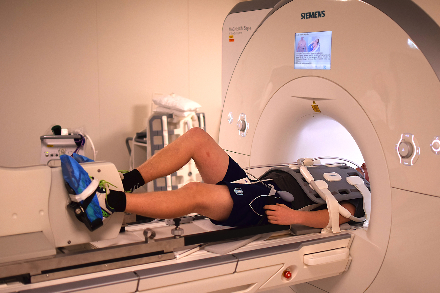
The method developed at the Göttingen Campus involves an ergometer similar to a home trainer, which is installed on the examination couch. The special feature is the MRI ergometer's non-magnetic components, which enable its use in the magnetic resonance tomograph's magnetic field. The patients are cycling lying down while the MRI scanner measures the pumping function of the heart. The doctors can follow the images on a screen during the examination and precisely assess how well the patient's heart works. "We see in the MRI how the heart beats, how it fills up and empties again," explains the first author of the study, Dr. Sören Backhaus, Department of Cardiology and Pneumology at UMG. "With MRI, we can therefore directly measure the pathogenic changes in the heart and not just assess their consequences." In patients with HFpEF, this shows that the performance of the left atrium of the heart is impaired. The left atrium is the first chamber of the heart that oxygenated blood reaches after the lungs. "However, this impaired function often only becomes apparent when the patients are moving and thus putting a strain on the cardiovascular system," says Backhaus.
Multi-centre studies planned
The current gold standard diagnostic examination requires a catheter to be inserted into the heart and the pulmonary artery and measures the change in lung pressure while the patients are exercising. In patients with diastolic heart failure, blood backs up into the lungs under stress, causing the lung pressure to increase. The examination with a cardiac catheter is exact but expensive, stressful for the patients, and not easy to implement because they have to move with a catheter inserted in their hearts.
With the DZHK study, the Göttingen scientists validated their new MRI examination and proved that it works very well for the diagnosis of HFpEF and that the catheter examination can thus possibly be avoided. However, larger studies are needed before the technique can be routinely used in diagnostics. "Real-time MRI bicycle ergometry is a completely new diagnostic procedure for patients with diastolic heart failure. Next, we are planning a study involving several centres to check whether the method is beneficial for patients," says Schuster.
The publication is Paper of the Month | January 2021 of the DZHK.
Study: Cardiovascular magnetic resonance real-time exercise stress testing in heart failure with preserved ejection fraction HFpEF-stress-DZHK17
Original publication: Exercise-Stress Real-time Cardiac Magnetic Resonance Imaging for Non-Invasive Characterisation of Heart Failure with Preserved Ejection Fraction: The HFpEF Stress Trial.
Backhaus SJ, Lange T, George EF, Hellenkamp K, Gertz RJ, Billing M, Wachter R, Steinmetz M, Kutty S, Raaz U, Lotz J, Friede T, Uecker M, Hasenfuß G, Seidler T, Schuster A.
Circulation. 2021 Jan 21.
DOI: 10.1161/CIRCULATIONAHA.120.051542.
Contact: Christine Vollgraf, Press and Public Relations, German Center for Cardiovascular Research (DZHK), Tel.: 030 3465 529 02, presse(at)dzhk.de
Scientific contacts: Prof. Dr. Andreas Schuster, Consultant Cardiologist and Professor of Medicine, Head of Cardiac Imaging Department of Cardiology and Pneumology, Heart Center, University Medical Center Göttingen, andreas.schuster(at)med.uni-goettingen.de
PD Dr. Sören Backhaus, Department of Cardiology and Pneumology, Heart Center, University Medical Center Göttingen, soeren.backhaus(at)med.uni-goettingen.de
Background information: Real-time magnetic resonance imaging
A group of researchers at the Max Planck Institute for Biophysical Chemistry developed real-time MRI technology. The technology enables unprecedented temporal and spatial resolution of MRI imaging in real-time. The new technology developed by Professor Martin Uecker, Dr. Shuo Zhang, and Professor Jens Frahm can record the beating heart with a temporal resolution of 10 milliseconds, i.e., as an image series or MRI film with up to 100 images per second. Likewise, it can measure the blood flow in the human circulation in real-time and with high resolution.
In addition to the indication in the HFpEF-stress-DZHK17 to take MRI images while the subject is exercising, the method has other advantages: For example, for patients with cardiac arrhythmias or patients who cannot hold their breath for several seconds because of their condition. This also applies to children with congenital heart defects, for whom the previously required general anaesthesia can be shortened or even entirely omitted.
Joint and speech movements and swallowing processes can also be directly observed with real-time MRI, which means that causes of pain or disorders can be discovered. In addition to the University Medical Centre Göttingen, several other Universities in Germany, Great Britain, and the USA are testing the Göttingen real-time MRI technology for routine use on patients.
Real-time MRI films of the Frahm Research Group, Max Planck Institute for Biophysical Chemistry Göttingen
Publications: Frahm J, Voit D, Uecker M. Real-Time Magnetic Resonance Imaging: Radial Gradient-Echo Sequences With Nonlinear Inverse Reconstruction. Investigative Radiology 2019;54:757-766. doi: 10.1097/RLI.0000000000000584
Uecker M, Zhang S, Voit D, Karaus A, Merboldt K-D, Frahm J. Real-time magnetic resonance imaging at a resolution of 20 ms. NMR in Biomedicine 2010;23:986-994. https://doi.org/10.1002/nbm.1585
This scientific publication was selected as Paper of the Month January 2021 by the DZHK Board of Directors.