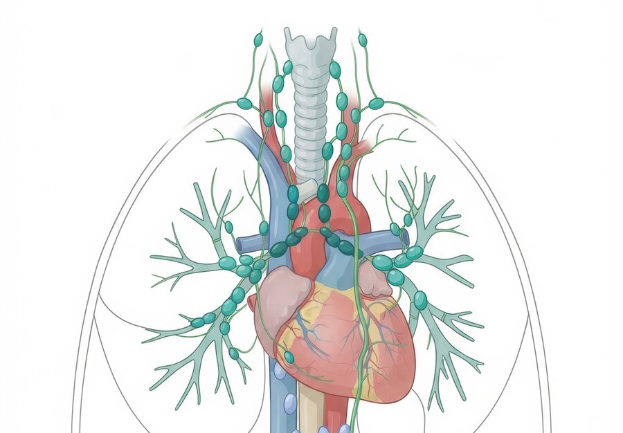
When people talk about the heart's vascular system, they usually mean the blood vessels. But the heart also contains lymphatic vessels: these drain excess fluid, proteins and cellular debris from the tissue and carry immune cells to the lymph nodes, helping to keep heart tissue healthy.
The team led by Professor Stefanie Dimmeler and Dr Julian Wagner at Frankfurt University Hospital investigated how the cardiac lymphatic system changes with age. They found significantly fewer lymphatic vessels in the hearts of older mice than in young animals, and made the same finding in human hearts. They also observed changes in the remaining lymphatic vessels: their endothelial cells were more tightly connected to one another, which impaired their ability to take up fluid and cells.
When the heart's drainage falters
The researchers observed that lymphatic vessels were already disappearing before typical age-related changes in the heart became apparent. Only later did immune cells, fluid, fibrinogen and amyloid begin to accumulate in the heart tissue. Cardiomyocytes enlarged, fibrosis increased, and diastolic function deteriorated — although systolic (pumping) function was preserved.
But do the missing lymphatic vessels actually cause these changes? To find out, the researchers deliberately reduced the number of lymphatic vessels in the hearts of young mice. Here too, macrophages and fibrinogen accumulated in the heart tissue, and diastolic function worsened, even though the heart continued to pump normally. The findings suggest that loss of lymphatic vessels accelerates cardiac ageing.
IL-33 drives the loss of lymphatic vessels
To pin down the underlying cause, the team examined various signalling molecules in the ageing heart. One signalling molecule in particular, interleukin-33 (IL-33), emerged as a key player. The researchers found increasing levels of IL-33 in lymphatic endothelial cells with advancing age. Cell culture experiments showed that this molecule inhibits the renewal of lymphatic vessels and promotes their degeneration.
One likely driver is the chronic, low-grade inflammation that increases with age. The researchers showed that inflammatory signals promote the production of IL-33 — setting up a potential vicious circle: inflammation damages the lymphatic vessels, immune cells are less effectively cleared from the heart tissue, accumulate there, and further fuel inflammation.
Can lymphatic vessels be restored?
In the animal model, this process could at least partly be reversed. When the researchers specifically lowered IL-33 levels in the lymphatic vessels of older mice, new lymphatic vessels began to form, and macrophage numbers in the heart tissue fell.
The study highlights that lymphatic vessels may play a previously underappreciated role in cardiac ageing. Whether this mechanism can ultimately be exploited to prevent or treat age-related heart disease remains to be shown by further research.
Original publication: Wagner, J.U.G., Gulshan, H., Sultan, I. et al. Age-associated loss of lymphatic vessels promotes cardiac inflammation. Nat Cardiovasc Res (2026). doi:10.1038/s44161-026-00870-y


