Palpitations when we are excited, a racing pulse during exercise, a pounding heart in stressful situations: the heartbeat constantly adapts to our surroundings and circumstances. For the heart to beat regularly, electrical signals in the myocardium must be precisely coordinated. In some people, a genetic variant disrupts this process: after each heartbeat, the myocardium takes longer to recover electrically. Under stress, this can trigger dangerous arrhythmias. One such condition is long QT syndrome type 2 (LQT2).
A research team at UMG has now shown that in LQT2, it is not only the cardiomyocytes that are electrically altered. Sympathetic neurons, which speed up the heart rate during physical or emotional stress, among other things, may also be hyperexcitable. Their activity may further increase the heart’s susceptibility to arrhythmias.
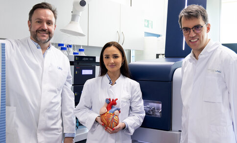
The team was led by Dr Maria-Patapia Zafeiriou, who has held the Else Kröner Professorship for Disease Modelling at UMG since June 2026, with support from Professor Wolfram-Hubertus Zimmermann, Director of the Institute of Pharmacology and Toxicology at UMG. The first authors of the study are Dr Lennart Valentin Schneider, a former doctoral student, and Dr Michael Gani Setya, a doctoral student at the Institute of Pharmacology and Toxicology at UMG.
The study in detail
In long QT syndrome, electrical recovery of the myocardium after each heartbeat is prolonged, which raises the risk of arrhythmias. The condition can have various genetic causes. In type 2 (LQT2), a potassium channel that helps to terminate electrical excitation in cardiomyocytes is impaired. Alterations in such channels can also affect neuronal function and, for example, promote epileptic seizures.
While the effects of the genetic variant on the myocardium are well characterised, its influence on autonomic neurons, which regulate heart rate among other functions, had until now remained unclear. Because emotional stress can trigger arrhythmias in LQT2, the researchers investigated whether the neurons that activate the heart under stress also contribute to their development.
Modelling heart and nerve cells
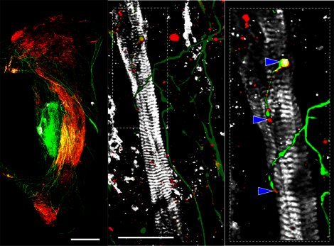
To address this question, the team led by Dr Schneider developed a human 3D model in which cardiomyocytes and sympathetic neurons are connected. They derived both cell types from human stem cells and used them to engineer three-dimensional tissues.
The neurons extended fine nerve fibres that grew into the cardiac tissue and made contact with the cardiomyocytes. The researchers were also able to demonstrate that this connection was functional: when the neurons were selectively activated with light, the beating rate of the cardiac tissue changed.
The researchers then examined cardiomyocytes and neurons carrying altered potassium channels. This revealed that the sympathetic neurons showed increased electrical excitability. Even when combined with healthy cardiomyocytes, they were able to compromise the electrical stability of the cardiac tissue. These findings indicate that diseased neurons can themselves contribute to the development of arrhythmias.
Effect of drugs on electrical activity
The beta blocker propranolol was unable to prevent the aberrant activity triggered by neuronal activation. The drug mexiletine, by contrast, reduced it. Mexiletine blocks sodium channels and thereby influences the electrical excitability of both cardiomyocytes and neurons.
Beta blockers are part of the standard treatment for LQT2. However, they do not offer all patients sufficient protection against arrhythmias. The findings point to further mechanisms that may play a role in LQT2. Whether they could lead to new therapeutic approaches will need to be tested in further studies.
“The results provide a possible indication that, in arrhythmias caused by altered potassium channels, the electrical excitability of neurons should be considered alongside that of the myocardium,” says Professor Zafeiriou.
A new model for cardiac research
The new model makes it possible to study cardiac muscle and neurons together and to combine them in a targeted manner. This allows researchers to distinguish the respective contributions of altered cardiomyocytes and altered neurons to the development of arrhythmias.
“In future, the model could help us to better understand the interplay between the heart and the nervous system in other diseases, and to test new drugs in a human tissue model,” says Professor Zafeiriou.
Source: Press release UMG
Original publication:
Schneider LV et al. Innervated human cardiac muscle model reveals sympathetic drivers of KCNH2-associated arrhythmias. Nature Communications (2026). DOI: 10.1038/s41467-026-76956-9