
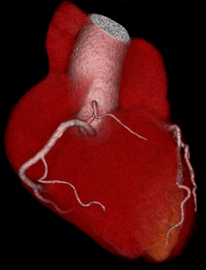
Quantitative imaging has become increasingly important for the diagnosis of coronary artery disease (CAD) over the past five years. This is because new quantitative techniques can detect narrowed coronary arteries (coronary artery stenoses) and atherosclerosis, which play a major role in CHD patients. It is important to correctly diagnose and accurately assess the severity of coronary artery stenosis or the extent of atherosclerotic burden for the selection of appropriate measures of therapy and the related further course of the disease. However, the complexity and variety of different quantitative imaging modalities, such as computed tomography (CT), magnetic resonance imaging (MRI), invasive coronary angiography (ICA), intravascular ultrasound (IVUS), and optical coherence tomography (OCT), necessitated a comprehensive clinical consensus.
Computed tomography is appropriate for suspected CAD with intermediate probability
Experts from numerous disciplines, including radiology, cardiology, and cardiac surgery, as well as engineering and computer science, have now compared the current status of different quantitative imaging methods for coronary artery stenosis and atherosclerosis in detail in a consensus paper, evaluating the potential of each method.
According to the authors, the clinical consensus clearly stated two things: First, noninvasive CT should be considered the preferred method in patients with intermediate probability of CAD to exclude or confirm narrowed or occluded cardiac vessels (obstructive stenoses). CT allows the assessment of deposits in the coronary arteries in terms of size, composition, location, and associated risk of future cardiovascular disease. Second, for patients presenting with acute symptoms or with a high probability of CAD, invasive methods such as ICA should be used preferred, including the possible addition of IVUS and OCT.
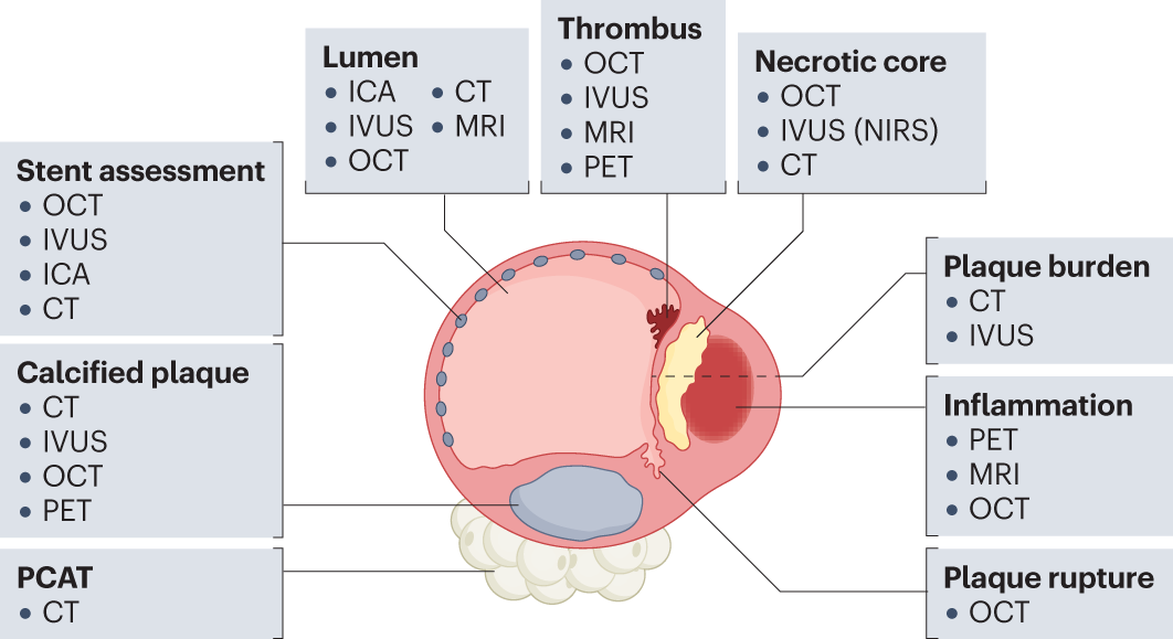
In addition, MRI can also facilitate the visualization of coronary plaques and serve as a radiation-free option for noninvasive coronary angiography in experienced centers. Positron emission tomography (PET) has the greatest potential for quantifying inflammation in coronary plaque. Although ICA is considered by the authors to be the reference standard for the assessment of stenosis, the method does not allow the characterization of coronary plaques. For plaques at high risk of rupture, IVUS and OCT are considered the main invasive imaging modalities for identification.
Quantitative imaging allows for more objective diagnosis and assessment of treatment progress
Prof. Dr. Marc Dewey, last author of the study expects the publication to change clinical practice, "This paper provides an interdisciplinary clinical consensus based on evidence-based data. It is a condensed summary and clearly maps current knowledge of each quantitative cardiovascular imaging procedure. Clinicians can thus be assisted in selecting the most appropriate method based on the specific clinical setting, individual patient characteristics, and availability of each procedure."
A total of seven methods were compared: Computed Tomography (CT), Magnetic Resonance Imaging (MRI), Positron Emission Tomography (PET), Single Photon Emission Computed Tomography (SPECT), Invasive Coronary Angiography (ICA), Intravascular Ultrasound (IVUS), and Optical Coherence Tomography (OCT). The advantage of quantitative imaging over conventional diagnostics, which only visually displays the acquired images, is the additional acquisition of biophysical parameters. This leads to a more objective diagnosis and allows an assessment of the course of therapy. In biomedical research and clinical diagnostics, quantitative imaging thus enables changes in specific tissues of the body to be determined.
The publication was made possible through funding from the German Research Foundation (DFG) for the Second Quantitative Cardiovascular Imaging Meeting (DE 1361/22-1).
Original publication: Mézquita AJV, Biavati F, Falk V, Alkadhi H, Hajhosseiny R, Maurovich-Horvat P, Manka R, Kozerke S, Stuber M, Derlin T, Channon KM, Išgum I, Coenen A, Foellmer B, Dey D, Volleberg RHJA, Meinel FG, Dweck MR, Piek JJ, van de Hoef T, Landmesser U, Guagliumi G, Giannopoulos AA, Botnar RM, Khamis R, Williams MC, Newby DE, Dewey M. Clinical quantitative coronary artery stenosis and coronary atherosclerosis imaging: a Consensus Statement from the Quantitative Cardiovascular Imaging Study Group. Nat Rev Cardiol. 2023 Jun 5. https://doi.org/10.1038/s41569-023-00880-4
Scientific contact: Prof. Dr. Marc Dewey, Heisenberg Professor of Noninvasive Cardiovascular Imaging and Vice Chair of the Department of Radiology at Campus Charité Mitte, Charité – Universitätsmedizin Berlin, marc.dewey(at)charite.de
