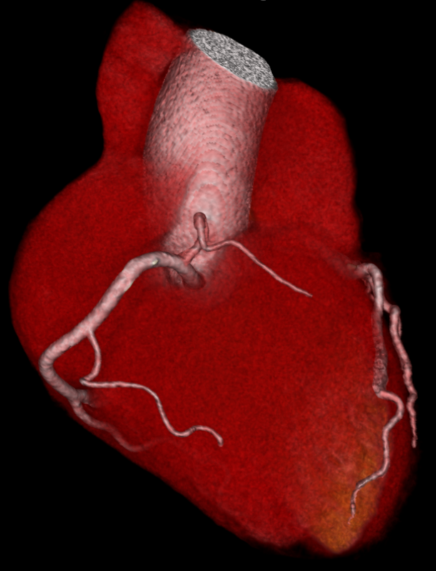
Coronary heart disease (CHD) is one of the most common heart diseases. In Germany alone, more than five million people are affected. In most cases, CHD is caused by calcification of the arteries, known as arteriosclerosis. The deposits cause the blood vessels that supply the heart to constantly narrow. Initially, symptoms such as chest pain and shortness of breath occur during physical exertion. If left untreated, blood clots can form from the deposits, which can completely block the vessel and lead to a heart attack.
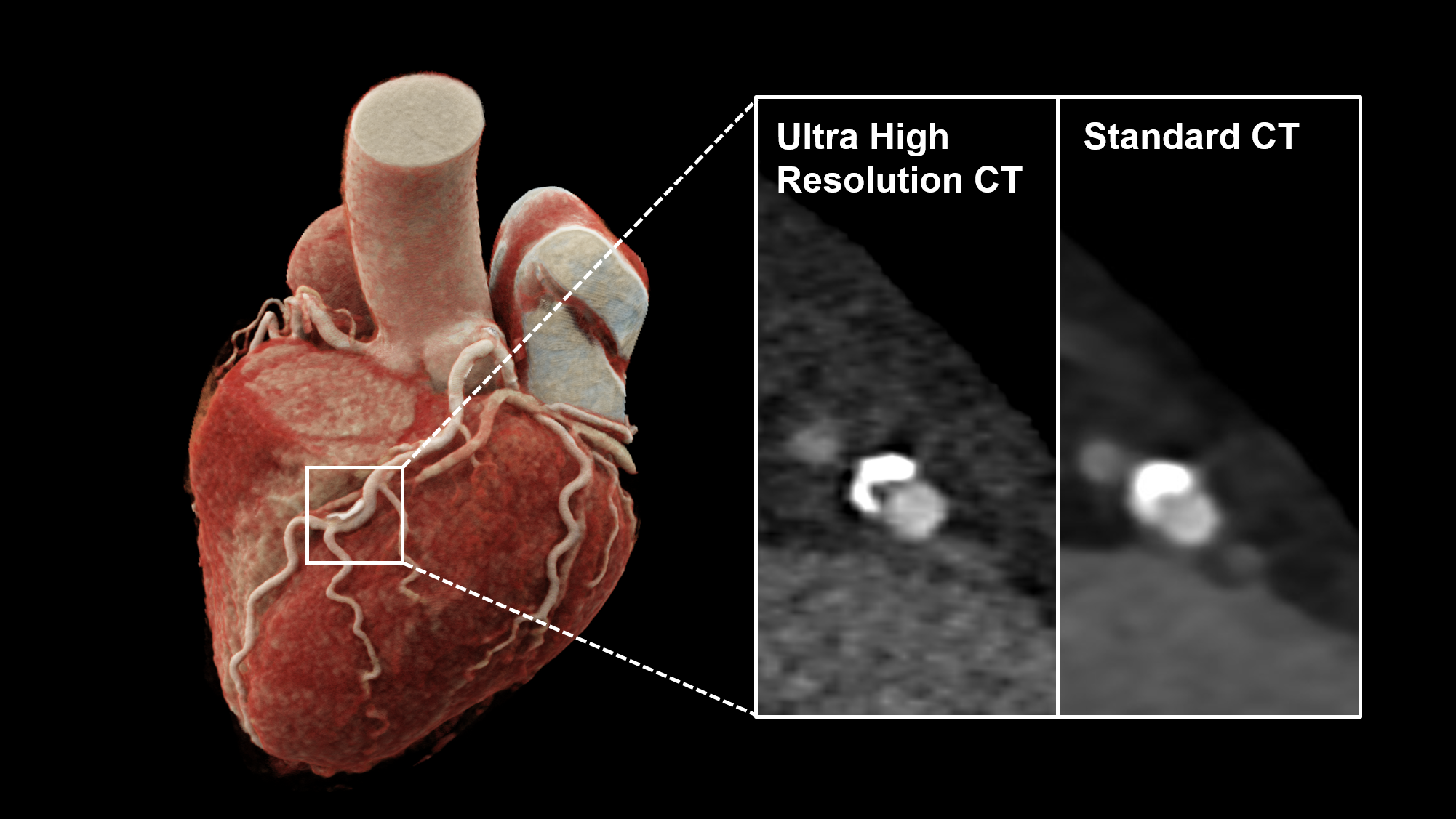
A heart examination using computerised tomography is one of the diagnostic procedures used to assess coronary heart disease. However, this imaging method has so far reached its limits, particularly in patients in whom the deposits are already heavily calcified. "Calcified vascular deposits have a higher density and appear more serious than they actually are due to the so-called calcium-blooming effect in cardiac CT. This can lead to an overestimation of the vasoconstriction, i.e. the stenosis," explains Dr Tilman Emrich, Senior Physician at the Clinic and Polyclinic for Diagnostic and Interventional Radiology at the University Medical Center Mainz and Assistant Professor of Radiology at the Medical University of South Carolina in Charleston.
Better image quality and spatial resolution
A new generation of computerised tomography scanners, known as photon counting detector CTs (PCD-CT), have significantly improved image quality compared to conventional CT. They also offer better spatial resolution. This means that two neighbouring structures, such as the vessel and the deposits, can be distinguished more precisely. "The new technology could be a significant advantage for patients whose stenosis has been overestimated due to the blooming effect. The improved assessment of coronary heart disease may significantly change the recommendations for downstream tests. This can potentially reduce unnecessary invasive procedures and lower healthcare costs," says Emrich.
In their study, the interdisciplinary cardiological-radiological research group led by Dr Tilman Emrich (Radiology) and Prof Dr Michaela Hell (Cardiology) examined 114 patients with suspected or diagnosed coronary heart disease using PCD-CT. They found that in many cases, ultra-high-resolution CT revealed a lower degree of stenosis than conventional CT. With a standard resolution, the degree of stenosis measured by the experts was significantly greater with a narrowing of 42 per cent than with the ultra-high resolution of 29 per cent. With the help of PCD-CT, around 54 per cent of the study participants could be classified in a lower stenosis category, the so-called CAD-RADS (Coronary Artery Disease Reporting and Data System) class, than they had originally been assigned. The effect was particularly large in people with severe vascular calcification. In mixed and non-calcified plaques with a low blooming effect, on the other hand, the scientists found no significant advantages of ultra-high resolution.
"In our study, we also investigated the effect of PCD-CT on an artificial vascular model. The model simulated a vessel with calcified deposits corresponding to a degree of stenosis of 25 and 50 per cent. The advantage of the ultra-high spatial resolution was also evident here. The reconstruction of the scans with the ultra-high resolution deviated from the model value by only around two to three per cent. With the standard resolution, the deviation was around 10 per cent," explains Dr Moritz Halfmann, first author of the publication and assistant physician at the Clinic and Polyclinic for Diagnostic and Interventional Radiology at the University Medical Center Mainz.
Potential for optimised diagnosis and treatment
"In our study, the innovative CT method shows clear potential to take a further step towards optimised and patient-centred diagnosis and treatment of coronary heart disease and to promote interdisciplinary collaboration between imaging and clinical cardiology experts. However, as our investigation is a simulation study, further validation of the results in comparative studies is required first," emphasises Professor Hell, Senior Physician at the Center for Cardiology I.
In computed tomography, energised particles of X-rays penetrate the tissue to be examined. The particles lose energy in the process, depending on how dense or permeable the tissue is. A special detector measures the remaining particle energy and converts it into electrical signals from which an image of the tissue is calculated. Previous detectors require an additional conversion step to generate this electrical signal from the measured energy. The standard detectors combine the energy of several particles and detailed information is lost. The new photon counting detector, on the other hand, can convert the individual particle energy directly into an electrical signal. As a result, each signal is converted so that the CT images have a higher spatial resolution and better quality.
Original publication: Ultra-High-Spatial-Resolution Photon-counting Detector CT Angiography of Coronary Artery Disease for Stenosis Assessment (Halfmann et al., 2024, Radiology)
Scientific contact: Dr Tilman Emrich (tilman.emrich(at)unimedizin-mainz.de), Clinic and Polyclinic for Diagnostic and Interventional Radiology, University Medical Center Mainz
Source: JGU press release (in German only)